Note: The upper T-spine may not be visible on the lateral view - if injury is suspected here then a swimmer's view may be helpful - (see Cervical spine - Normal). Images of the thoracic and lumbar spine are often large and the bones should be scrutinised in detail (see images below). Thoracic spine - Standard viewsĪP and Lateral - Assess both views systematically (see box). The clinico-radiological assessment of suspected T-spine or L-spine injuries therefore depends on careful consideration of both the clinical and radiological findings. Clinical assessment is also often limited by distracting injuries or reduced consciousness. Good views of the T-spine and L-spine are difficult to achieve in the context of trauma. Imaging should not delay resuscitation.įurther imaging with CT or MRI (not discussed) is often appropriate in the context of a high risk injury, neurological deficit, limited clinical examination, or where there are unclear X-ray findings. Therefore, patients with suspected spinal injury should be managed by experienced clinicians in accordance with local and national clinical guidelines. Incorrect management of patients with spinal injury may cause or worsen neurological deficit. The plain X-ray anatomy and appearances of injuries to both these areas are discussed together. One of these methods is Chiropractic BioPhysics (CBP) technique which is a full-spine and posture treatment that utilizes mirror image (i.e. In the context of trauma similar principles apply to imaging both the Thoracic spine (T-spine) and the Lumbar spine (L-spine). Spacing - Discs/Spinous processes/Pedicles.Bones - Cortical outline/Vertebral body height.Thoracolumbar spine - Systematic approach If you see one fracture - check for another.1 Specifically, these degenerative changes may lead to the formation of bone spurs called osteophytes. Nerve root encroachment is often caused by degenerative ('wear and tear') changes in the vertebrae, which is part of the normal aging process. If 'instability' is suspected then further imaging with CT should be considered When you have nerve root encroachment, abnormal tissue moves in on the spinal nerve root.Huge collection, amazing choice, 100+ million high quality, affordable RF and RM images. Correlate radiological findings with the clinical features Find the perfect align your spine stock photo.

It’s not just coincidence that people most with poor posture tend to lack energy and suffer from fatigue, experience random aches and pains, as well as suffer from headaches / migraines, allergies, ear infection, sleeping disorders, gastrointestinal issues (such as GERD), asthma, high blood pressure, etc.Īs a CBP patient, you will notice not only a dramatic decrease in pain in discomfort, but more importantly a sense of restored health and energy.

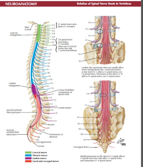
Thus, your organs are much more susceptible to malfunction and disease. Just as a kink in a garden hose can dramatically reduce the flow of water coming from the tap – a spine out of alignment dramatically reduces vital information going from your brain to vital organs. From your immune, cardiovascular, and digestive system, to sexual function and mobility – your nerves are responsible for sending vital information and energy from the brain to your organs through your spine.


Your spine houses your nervous system – the most delicate and important organ system – responsible for your day to day bodily functions.


 0 kommentar(er)
